Why technique matters in otoplasty
Otoplasty, sometimes called ear pinning, is surgery that changes the position, shape or proportion of the outer ear. The visible ear is made largely of elastic cartilage covered by skin, so the operation is not simply a matter of pulling the ear backward. A surgeon first studies the ear’s folds, rim, conchal bowl, skin quality, symmetry and relationship to the side of the head. The plan may address one feature or several features together.
The phrase “ear pinning” can therefore describe different surgical actions. One plan may create a more defined antihelical fold, another may reduce a prominent conchal area, and another may combine cartilage reshaping with carefully placed sutures. Some procedures use an incision behind the ear; selected approaches may use needle access or small openings. The suitable route depends on anatomy, age, cartilage flexibility, previous injury or surgery, and the change being discussed. There is no single technique that is appropriate for every ear.
**Medical notice:** This article is general education, not a diagnosis or a personal treatment plan. Otoplasty technique, anesthesia, timing, risks and aftercare must be assessed by a qualified surgeon after an examination. Do not use this information to decide whether surgery is suitable for you.
The anatomy a surgeon may assess
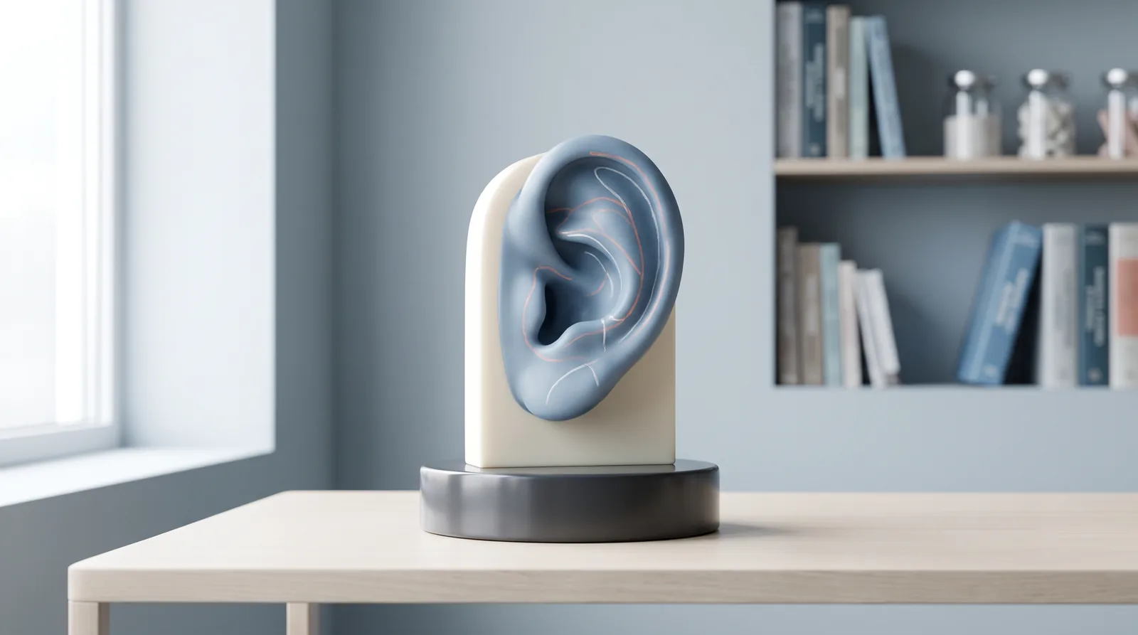
The outer ear has landmarks that help create a balanced result. The helix is the outer rim. The antihelix is an inner curved fold that can be less defined in a prominent ear. The concha is the deeper bowl next to the ear canal. The lobule is the soft lower portion and contains little cartilage. During consultation, the surgeon may compare the two ears from the front, side and back, noting differences that are normal and cannot always be made identical.
Assessment can also include skin thickness, cartilage strength and spring, the angle between the ear and skull, and the position of the hairline. A child’s cartilage may be more flexible than an adult’s, while previous trauma can make the tissue irregular or scarred. The surgeon may ask about ear infections, allergies, medicines, smoking, bleeding problems and previous operations. These details can influence whether a technique is considered and how recovery is planned.
Cartilage-shaping approaches
Creating or reinforcing an antihelical fold
When the inner fold is shallow, a surgeon may form or reinforce it with sutures placed through the cartilage from an incision, usually behind the ear. The sutures bend the cartilage along a planned curve. The aim is not to create a sharp or identical fold in every person; the curve must suit the ear’s natural thickness and the opposite side. Over-tightening can create an overly rigid or artificial contour, which is why placement and tension require judgment.
Modifying the conchal cartilage
A deep or prominent conchal bowl can contribute to the ear sitting farther from the head. Depending on the anatomy, the surgeon may use sutures to set the conchal cartilage back toward the mastoid area, remove a limited amount of cartilage, or use a combination. Removing tissue is not automatically necessary. Preserving adequate support matters because excessive reduction can alter the ear’s contour or create unwanted irregularity.
Scoring, weakening or reshaping cartilage
Cartilage can have a natural tendency to return toward its original curve. Some methods use controlled scoring, rasping or weakening of selected cartilage surfaces so the tissue bends more easily. Other methods rely mainly on sutures. These approaches can be combined, but they have different implications for contour, stability, palpable edges and scar placement. The surgeon should explain what is being changed and why that step fits the examined anatomy.
Suture-focused or limited-access methods
Some otoplasty plans use a needle or small access points to place sutures without a long skin incision. The needle may make the cartilage more flexible, while sutures hold the ear in a revised position or connect it to tissue behind the ear. These approaches may not suit cartilage that is very stiff, thick, misshapen or affected by prior surgery. “Less visible incision” does not mean no risks, no marks or guaranteed durability. A consultation should clarify how the surgeon would manage asymmetry, suture visibility, recurrence or a need for revision.
Where incisions may be placed
A common open approach uses an incision in the crease behind the ear. Through this access, the surgeon can see the cartilage, place internal sutures and remove a small amount of tissue when indicated. The skin incision is then closed, and the scar may settle within the natural shadow behind the ear. The final appearance of a scar varies with healing, skin characteristics and aftercare.
If access to the front surface is needed, incisions may be placed within natural folds so they are less conspicuous. The choice is not based only on hiding a line. It must provide enough visibility and control to reshape the specific structures safely. A plan should also consider hair follicles, skin tension, blood supply and the possibility that a front-surface mark could be visible in some lighting.
“Incisionless” or limited-incision descriptions need careful explanation. A technique may avoid a traditional skin cut while still using needles, cartilage manipulation and permanent or long-lasting sutures. It may be useful in selected anatomy, but it is not automatically safer or more appropriate. Ask what access is proposed, which tissues will be altered, and how the surgeon will monitor the ear during healing.
How sutures help position the ear
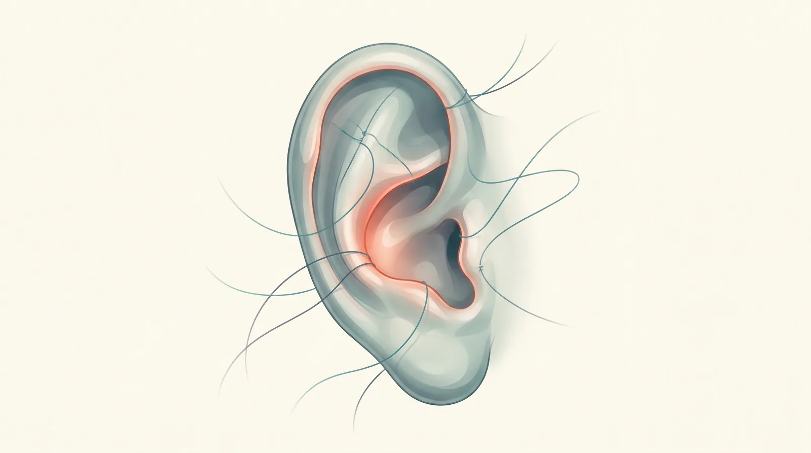
Internal sutures can create a fold, draw the ear closer to the head, or secure cartilage to a stable tissue layer behind the ear. They may be permanent or absorbable depending on the surgeon’s method. External stitches close the skin and may be removable or dissolvable. The number and location of sutures vary; a higher number does not by itself indicate a better plan.
Suture tension is important. Too little support may allow a fold to relax, while excessive tension can cause discomfort, visible puckering, cartilage pressure or an unnatural contour. Suture-related issues can include palpability, extrusion, infection or later loosening. These are uncommon but should be included in informed consent. A surgeon should describe how such problems would be evaluated and managed rather than promising that they cannot occur.
Dressings, healing and activity
After surgery, a dressing or head bandage may support the new position and protect the ears. It should be kept clean and dry and changed only according to the clinical instructions. Some patients are advised to use a soft headband at night for a period of time to reduce accidental pressure during sleep. Button-front clothing can help avoid catching the ears while dressing.
Tenderness, swelling, bruising, itching, tingling or temporary numbness can occur. Recovery is individual. A follow-up is often arranged within about a week, with additional visits as needed. Return to school or desk work may occur relatively soon for some people, while strenuous activity, swimming and contact sports usually require longer restrictions. The exact schedule depends on the operation and the surgeon’s assessment, not on a universal calendar.
Seek prompt medical advice for increasing pain, marked one-sided swelling, excessive bleeding, fever, spreading redness, drainage, an injury to the surgical area or any symptom that feels unexpected. A collection of blood near the cartilage and infection are among the complications clinicians watch for. Asymmetry, scar concerns, stiffness, altered sensation or partial return of prominence may also occur. Early review can matter when a problem is suspected.
Questions for an individual consultation
A useful consultation is a conversation about anatomy rather than a choice between attractive procedure names. Ask which landmarks are contributing to prominence, whether the plan involves cartilage scoring, cartilage removal, sutures, fixation behind the ear, or a combination, and where each incision would be. Ask whether sutures are intended to remain, what the dressing and activity restrictions will be, and how follow-up is arranged.
It is also reasonable to ask about anesthesia, medicines that may increase bleeding, smoking or nicotine, medical conditions, previous ear surgery and the surgeon’s approach to asymmetry or recurrence. Bring a list of medicines and supplements. For a child, discuss readiness, school arrangements and how to prevent pressure or impact during healing. The goal is informed consent based on the examined ear, not a promise of a fixed shape.
For a personal discussion of anatomy and options, Book an Otoplasty Consultation.
To place the discussion of techniques within the full treatment journey, explore the Otoplasty procedure page. You may also see the Otoplasty Before & After gallery when discussing anatomy and realistic goals. Images show individual clinical cases and do not determine which technique is suitable for another person.
Frequently Asked Questions
1. Is otoplasty always performed through an incision behind the ear?
No. A behind-the-ear incision is common, but some plans use incisions within natural folds or limited-access methods. The choice depends on the structures that need correction and the surgeon’s ability to work safely and precisely.
2. Does cartilage have to be removed during ear pinning?
Not always. Some ears can be repositioned with sutures or controlled cartilage reshaping. Small cartilage reduction may be discussed when the conchal area contributes to prominence, but the decision requires examination.
3. When can normal activities resume?
Light daily activity may return earlier than swimming, strenuous exercise or contact sports. Healing varies, so follow the individualized instructions and obtain clearance at follow-up rather than relying on a fixed date.
A measured way to understand technique
Otoplasty is a structural operation with several possible components. Cartilage may be folded, weakened, reduced or supported with sutures; incisions may be hidden behind the ear or placed in natural creases; and dressings help protect the early healing position. These choices are linked to the ear’s anatomy and the patient’s circumstances. Understanding the options can make consultation more productive, but only an examination can determine which discussion is relevant.