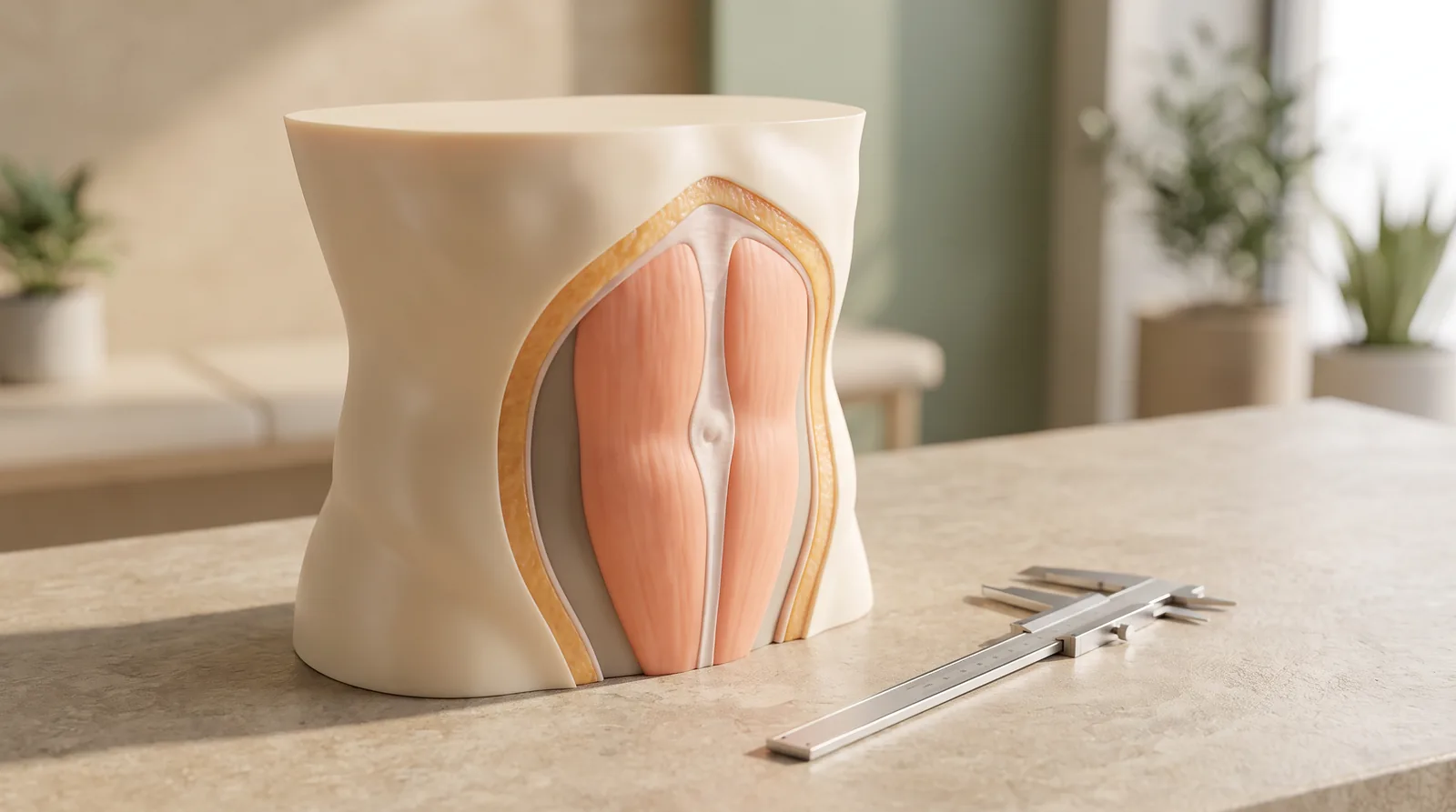
A lower-abdominal fullness is often described as “stubborn fat,” especially after pregnancy, weight change or a period away from exercise. That description can be understandable, but it is not a diagnosis. The visible contour of the abdomen comes from several layers: skin, subcutaneous fat, the abdominal wall and the tissues deeper inside the abdomen. Diastasis recti is a widening of the connective tissue in the midline between the two rectus abdominis muscles. It can change the way the abdominal wall manages pressure and may produce a central dome during effort. Fat reduction, by contrast, addresses deposits in the layer beneath the skin. They are different concerns, so a responsible abdominal-contouring consultation starts by identifying which layer, or combination of layers, is contributing to the concern.
Why a forward abdomen needs a layered assessment
A contour concern is not reliably identified by the word “bulge.” A soft, pinchable layer that is relatively consistent from the upper to lower abdomen may suggest a meaningful subcutaneous-fat component. Loose or creased skin may be a separate issue, particularly after weight loss or pregnancy. A central ridge or dome that becomes more apparent when rising, coughing or bracing can prompt an assessment of the abdominal wall. These patterns may coexist in the same person; none of them should be assumed from a photograph, a scale reading or a single measurement.
Diastasis recti refers to the midline connective tissue, called the linea alba, becoming widened and thinned as the paired rectus muscles move apart. Pregnancy is a common setting, but it is not the only one. Changes in abdominal pressure, body weight, training history and the tissues’ individual response can all be relevant. Some people notice an altered shape only. Others describe difficulty generating a comfortable sense of core support during certain tasks. A gap size alone does not tell the whole story: the quality of tension across the midline, symptoms, daily function and the person’s goals all matter.
A hernia is not the same as diastasis. With diastasis, the midline tissue is stretched rather than a true hole in the abdominal wall. A hernia involves a defect through which tissue may protrude. They can occur together, which is one reason a clinician should assess a new or focal bulge rather than assuming it is simply fat or muscle separation.
How suspected diastasis is assessed in clinic
Assessment normally begins with a conversation, not with a treatment plan. A clinician may ask about pregnancies, abdominal operations, major weight changes, constipation or repeated straining, exercise and work demands, discomfort, urinary or pelvic-floor symptoms, and the specific moments when the contour changes. The purpose is not to judge the abdomen; it is to understand the pattern and to screen for concerns that may need a different referral or investigation.
The physical examination commonly looks at the abdomen while relaxed and while gently engaging the trunk. Observation can be useful in standing and lying positions because gravity, posture and muscle recruitment change the contour. The clinician may feel along the midline above, at and below the navel to estimate the separation and assess the resistance of the tissue. How the abdominal wall behaves during a controlled movement can be more informative than how it looks at rest. Visible doming is a finding to interpret in context, not proof of a particular severity.
Measurement may be documented so that the discussion is consistent over time. Clinical examination and finger-width estimation can help identify a separation; ultrasound or calipers may be used when a more precise inter-rectus measurement would add value. Imaging is not an automatic requirement for every contour concern. It may be considered when the examination is unclear, a hernia is suspected, or it would help surgical planning. A clinician also considers skin quality, fat distribution, scars, posture and signs that the fullness may arise deeper inside the abdomen rather than from a superficial contour layer.
This is different from an assessment focused mainly on localized fat. For that question, the distribution and pinch of subcutaneous tissue, skin recoil, the person’s weight stability and the relationship between the area of concern and the rest of the torso are central. The consultation still includes general health, medications, smoking or nicotine exposure, prior procedures and expectations. It does not use the amount of fat as a shortcut for diagnosing diastasis.
Why fat reduction and diastasis repair are different decisions
Fat reduction procedures are designed to change selected deposits of subcutaneous fat. They do not bring separated rectus muscles closer together, restore tension to a widened linea alba or repair a hernia. They also cannot predictably correct loose skin. Conversely, an abdominal-wall repair is aimed at the supporting layer and does not automatically address every contour feature. If excess skin or fat is also present, that is a separate part of the discussion and may or may not be appropriate to combine with another procedure.
This distinction matters because the same outward shape can have different drivers. A person with a relatively small amount of pinchable fat may still show midline doming under effort. Another person may have abdominal fullness primarily related to superficial fat, with no clinically significant diastasis. A third may have a mix of skin laxity, fat distribution and altered abdominal-wall support. Seeking “fat removal” when the main issue is dynamic midline support can leave the original concern unaddressed. Seeking muscle repair when the main concern is fat or skin can be equally mismatched.
Where diastasis is present, non-operative care may be discussed before any procedure. Individualized guidance from a suitably trained clinician or physiotherapist can focus on pressure management, trunk coordination and movement choices. It is important not to treat a generic online exercise list as a prescription. A movement that causes marked doming, pain or a sense of strain deserves review, and the correct progression depends on the person’s circumstances. Improvement in function or control does not require that every measurement become identical, and the evidence does not support promising a specific outcome from a single approach.
If surgery is considered, shared decision-making should be specific about the anatomical target. Repair of diastasis may involve tightening the midline supporting tissue; the plan, approach and whether another contouring element is considered depend on examination, health factors and the presence or absence of a hernia. Future pregnancy, major anticipated weight changes, medical conditions and previous scars can influence timing and suitability. An ethical consultation explains trade-offs, scars, anaesthesia and recovery demands as well as potential benefits; it does not present a flatter abdomen as a guaranteed result.
Treatment boundaries, safety and informed choice
Abdominal contouring is not a substitute for weight management, treatment of digestive symptoms or diagnosis of a new abdominal mass. It is also not appropriate to assume that back pain, pelvic symptoms or urinary leakage are caused by diastasis alone. These concerns may need assessment by the relevant healthcare professional. An abdominal binder may offer temporary support for some situations, but it does not repair the linea alba or replace an evaluation.
Safety assessment includes more than the abdomen. It includes medical history, prior operations, medication and supplement use, nicotine exposure, clotting or wound-healing risks, plans for pregnancy, and whether goals are realistic for the tissue present. A sudden painful or tender bulge, skin colour change over a bulge, vomiting, fever, or an abdominal swelling that cannot be gently reduced should be evaluated promptly, as these symptoms may be consistent with a hernia complication or another acute condition.
No article can determine whether a person has diastasis or which option is suitable. A clinical assessment may conclude that reassurance, rehabilitation, medical evaluation, a staged approach or no procedure is the safest path. The best outcome is not defined only by a measurement or a photograph; it should include an informed understanding of what the selected approach can and cannot change.
Frequently Asked Questions ### Can liposuction diagnose or fix diastasis recti?
No. Liposuction is not a diagnostic test for the abdominal wall, and fat reduction does not repair a widened linea alba or a hernia. If there is concern about a central bulge or dynamic doming, that should be examined before deciding whether fat reduction is relevant.
If the gap seems small, can it still matter?
The width is only one finding. Clinicians also consider location, tissue tension, core control, symptoms and whether a hernia is present. Equally, a wider measurement does not by itself establish the need for surgery. The interpretation is individualized.
Should I avoid all abdominal exercise if I notice doming?
Do not rely on a blanket rule. Pain, pronounced bulging or uncertainty about technique are good reasons to seek professional advice. A qualified clinician or physiotherapist can assess movement and recommend an appropriate, gradual plan rather than a one-size-fits-all routine.
**Medical notice:** This article provides general education and is not a diagnosis, examination or personal treatment recommendation. Seek urgent medical care for severe or worsening abdominal pain, persistent vomiting, fever, or a painful non-reducible bulge.
For a clinician-led discussion of abdominal contour concerns and their anatomical causes, visit Body Contouring.